Home
/ Back Of Neck Anatomy - Neck Pain - OrthoInfo - AAOS / Foundational anatomy provides medical students with the necessary background in anatomy for success in clerkships.
Back Of Neck Anatomy - Neck Pain - OrthoInfo - AAOS / Foundational anatomy provides medical students with the necessary background in anatomy for success in clerkships.
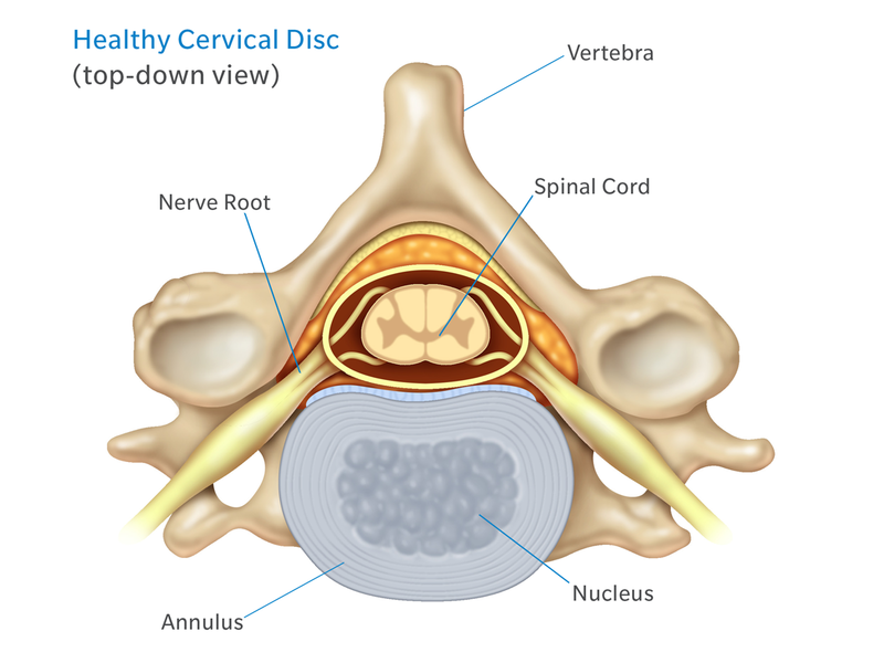
Back Of Neck Anatomy - Neck Pain - OrthoInfo - AAOS / Foundational anatomy provides medical students with the necessary background in anatomy for success in clerkships.. The posterior muscles of the neck are primarily concerned with head movements, like extension. The back muscles stabilize and move the vertebral column, and are grouped according to the lengths and direction of the fascicles. Some important structures contained in or passing through the neck include the seven cervical vertebrae and enclosed spinal cord, the jugular veins and carotid arteries, part of the esophagus, the larynx. Clinically, surface anatomy is used to split the neck into anterior and posterior triangles which provide clues as to the location of specific structures. Muscles of the posterior neck and the back.
In radiology, the 'head and neck' refers to all the anatomical structures in this region excluding the central nervous system, that is, the brain and spinal co. Head and neck anatomy focuses on the structures of the head and neck of the human body, including the brain, bones, muscles, blood vessels, nerves in a newborn, the junction of the paritial bones with the frontal and occipital bones, form the anterior (front) and posterior (back) fontanelle, or soft spots. This article describes the anatomy of the head and neck of the human body, including the brain, bones, muscles, blood vessels, nerves, glands, nose, mouth, teeth, tongue, and throat. Your neck is like no other part of the vertebral spinal column and enables your head and neck a wide range of motion. This mri neck axial cross sectional anatomy tool is absolutely free to use.
Anatomy of The Neck: Causes of Neck Pain and How to Manage ... from www.cervicaldisc.com Want to learn more about it? The back muscles stabilize and move the vertebral column, and are grouped according to the lengths and direction of the fascicles. Use the mouse scroll wheel to move the images up and down alternatively use the tiny arrows (>>) on both side of the image to move the images. The neck is an extremely complicated place in the body. Some important structures contained in or passing through the neck include the seven cervical vertebrae and enclosed spinal cord, the jugular veins and carotid arteries, part of the esophagus, the larynx. The spine runs from the base of your skull down the length of your back, going all the way down to your pelvis. Dummies helps everyone be more knowledgeable and confident in applying what they know. It provides images in the axial and coronal planes so that the user can study and learn anatomy.
Neck muscles help support the cervical spine and contribute to movements of the head, neck, upper back, and shoulders.
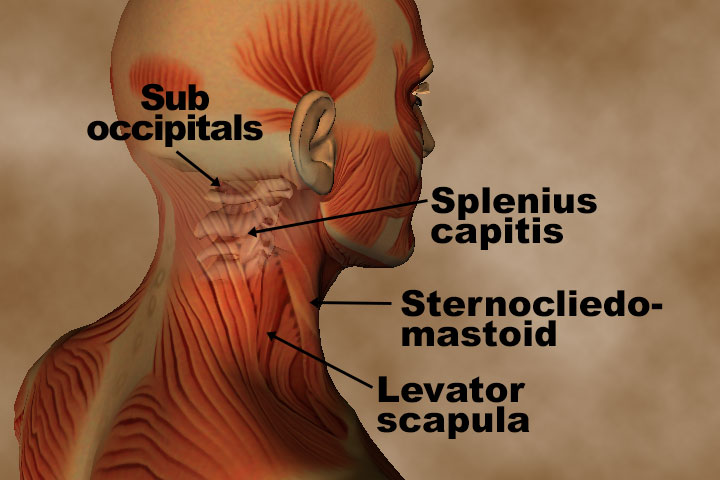
The cervical spine protects the nerves connecting to the brain, allowing the head to move freely while supporting its weight. It runs from the neck to the upper back. Some important structures contained in or passing through the neck include the seven cervical vertebrae and enclosed spinal cord, the jugular veins and carotid arteries, part of the esophagus, the larynx. The levator scapulae muscle is attached at the top four cervical vertebrae (c1 to c4) and runs down the side of the neck to attach at the top of the shoulder blade (scapula). We use cookies to ensure that we give you the best ex. They control the scapulae (shoulder blades), which play a role in shrugging, neck movement, head. The head rests on the top part of the vertebral column, with the skull joining at c1. Despite being a relatively small region, it contains a range of important anatomical features. It not only supports the brain in its quest against gravity, but supplies the passageways from this makes the neck a sort of nervous mecca for the entire body, it is the portal of the entire human body into the organs, through the mouth and also. Posterior triangle of the neck boundari… pretracheal fascia b. In radiology, the 'head and neck' refers to all the anatomical structures in this region excluding the central nervous system, that is, the brain and spinal co. 12 photos of the anatomy of the back of the neck. When most people mention their back, what they are actually referring to is their spine.
Learn about these muscles, their locations & functional the traps are quite a complex set of muscles. Understanding the anatomy of your cervical spine and the vital nerves it contains should motivate you to adopt behaviors that help prevent neck injury and. Use the mouse scroll wheel to move the images up and down alternatively use the tiny arrows (>>) on both side of the image to move the images. Our neck is where we find the seven cervical vertebrae, with c7 (the seventh cervical vertebra) meeting t1 (the first thoracic vertebra) at the base of the neck. Cervical spine anatomy video the cervical spine has 7.
Cardiovascular System of the Head and Neck from www.innerbody.com It is made up of bones, discs the neck is connected to the upper back through a series of seven vertebral segments. The back muscles stabilize and move the vertebral column, and are grouped according to the lengths and direction of the fascicles. The head rests on the top part of the vertebral column, with the skull joining at c1. This atlas is a comprehensive and affordable learning tool for residents and medical students and especially for radiologists and surgeons. Posterior triangle of the neck boundari… pretracheal fascia b. Head and neck anatomy is important when considering pathology affecting the same area. 3d interactive tutorials on the anatomy of the neck, including the anatomical organisation, musculature, larynx, pharynx, blood supply and innervation. This entry was posted in anatomy by admin.
This article concerning the anatomy of the head and neck area gives you a clear structure at hand to see anatomy and function of the regions of the lower face.
The cervical spine protects the nerves connecting to the brain, allowing the head to move freely while supporting its weight. The neck is the area between the skull base and the clavicles. The neck is an extremely complicated place in the body. 12 photos of the anatomy of the back of the neck. Our neck is where we find the seven cervical vertebrae, with c7 (the seventh cervical vertebra) meeting t1 (the first thoracic vertebra) at the base of the neck. Foundational anatomy provides medical students with the necessary background in anatomy for success in clerkships. The back comprises the spine and spinal nerves, as well as several different muscle groups. How to view the anatomical labels. The neck is connected to the upper back through a series of seven vertebral segments. Cervical spine anatomy video the cervical spine has 7. Learn about these muscles, their locations & functional the traps are quite a complex set of muscles. This article concerning the anatomy of the head and neck area gives you a clear structure at hand to see anatomy and function of the regions of the lower face. They control the scapulae (shoulder blades), which play a role in shrugging, neck movement, head.
Anatomy of the head and neck: Choose from 500 different sets of flashcards about neck anatomy back neck upper on quizlet. We've largely focused on the physical aspect of our spinal anatomy in this series. Some important structures contained in or passing through the neck include the seven cervical vertebrae and enclosed spinal cord, the jugular veins and carotid arteries, part of the esophagus, the larynx. They control the scapulae (shoulder blades), which play a role in shrugging, neck movement, head.
Anatomy and Pathology for bodyworkers - Real Bodywork from www.realbodywork.com The anterior jugular vein (v. We use cookies to ensure that we give you the best ex. Despite being a relatively small region, it contains a range of important anatomical features. It consists of seven vertebrae. An overview of the anatomy of the hand, including the bones of the hand, muscles, blood supply and nerve supply. Head and neck anatomy focuses on the structures of the head and neck of the human body, including the brain, bones, muscles, blood vessels, nerves in a newborn, the junction of the paritial bones with the frontal and occipital bones, form the anterior (front) and posterior (back) fontanelle, or soft spots. The levator scapulae muscle is attached at the top four cervical vertebrae (c1 to c4) and runs down the side of the neck to attach at the top of the shoulder blade (scapula). The back comprises the spine and spinal nerves, as well as several different muscle groups.
The head rests on the top part of the vertebral column, with the skull joining at c1.
Head and neck anatomy is important when considering pathology affecting the same area. Clinically, surface anatomy is used to split the neck into anterior and posterior triangles which provide clues as to the location of specific structures. They control the scapulae (shoulder blades), which play a role in shrugging, neck movement, head. The neck is connected to the upper back through a series of seven vertebral segments. This atlas is a comprehensive and affordable learning tool for residents and medical students and especially for radiologists and surgeons. Learn everything about the neck anatomy with this topic page. The back muscles stabilize and move the vertebral column, and are grouped according to the lengths and direction of the fascicles. « back show on map ». When most people mention their back, what they are actually referring to is their spine. The posterior muscles of the neck are primarily concerned with head movements, like extension. This entry was posted in anatomy by admin. It consists of seven vertebrae. The head rests on the top part of the vertebral column, with the skull joining at c1.
